Treatment of the Atrophic Maxilla with Personalized Implants by CPMH: A Case Report
- Jan 23
- 2 min read
In contemporary implantology, one of the most significant challenges remains the aesthetic, phonetic, and functional reconstruction of jaws affected by severe bone loss. Such deficiencies typically result from severe atrophy or maxillectomies performed for the treatment of tumors or trauma.
Case Report
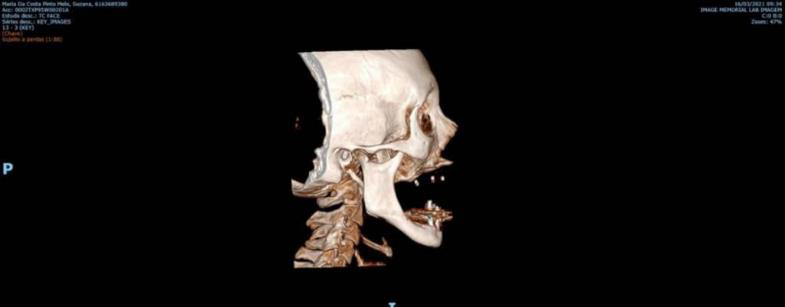
A female patient (S.M.C.P.M) presented to the clinic with a history of being totally edentulous in the maxilla for over 40 years. Her primary complaints were dissatisfaction with her current prosthesis, masticatory difficulties, and a profound sense of insecurity.Extraoral and intraoral clinical examinations (Figures 1, 2, and 3), supported by tomographic imaging (Figures 4 and 5), revealed substantial bone loss throughout the entire maxillary body bilaterally. This condition was attributed to long-term tooth loss and previous failed bone graft rehabilitation treatments.Imaging confirmed extensive pneumatization of both the right and left maxillary sinuses. Nearly total resorption of previous bone grafts was noted, with no signs of osseointegration from the initial treatment options proposed to the patient. Clinically, the patient was lucid and oriented with stable vital signs, though she reported hypertension and dysphagia (swallowing disorders), noting frequent choking on both liquid and solid foods.


Treatment Plan with CPMH CustomLife Reconstruction System
The proposed treatment plan involved rehabilitative surgery using a patient-matched maxillary prosthesis designed for implant-supported prosthetic rehabilitation. The objective was to restore maxillary anatomy and function, which had been compromised by advanced bone loss and previous surgical failures. Following preoperative evaluations and consultations, the patient was admitted to a private hospital in Salvador, State of Bahia, Brazil for the procedure.Once the digital surgical planning was approved (Figures 5 and 6), the procedure was performed under general anesthesia with nasotracheal intubation. The surgery began with an incision in the edentulous premaxilla, complemented by two vertical incisions in the region of the zygomatic pillars. Full-thickness mucoperiosteal flaps were reflected to define the surgical field.


Upon completion of the surgical site preparation, the CustomLife device was implanted following the predetermined digital stages. The procedure involved precise positioning of the patient-matched implant and verification of its anatomical alignment to ensure optimal adaptation and support for the subsequent prosthetic phase.

Results of treatment with superosteal implant by CPMH
The patient was monitored in a hospital setting for eight hours postoperatively. Finding no complications, she was provided with postoperative instructions and discharged.
Twenty-four hours after the surgery, the outpatient oral rehabilitation (prosthetic phase) commenced. The final prosthesis was installed three days post-surgery. Results were verified via panoramic radiography (Figure 8) and continuous clinical monitoring.

 |  |
Figure 9: Final clinical aspect following prosthesis installation
Personalized implants represent the next generation of maxillofacial surgery and implantology. They enable the installation of personalized prostheses for immediate-load rehabilitation without the need for bone grafts, significantly increasing the success rate of complex rehabilitative treatments.
Reference
DA MAIA, Renan Prado Reis et al. Treatment of atrophic maxilla with personalized implant: case report. Brazilian Journal of Health Review, v. 6, n. 4, p. 15904-15916, 2023. - https://doi.org/10.34119/bjhrv6n4-152


.png)
Comments